Anatomy Of Chest Wall / Surgical Anatomy Of The Chest Wall Springerlink / The chest wall, like other regional anatomy, is a remarkable fusion of form and function.
byAdmin-
0
Anatomy Of Chest Wall / Surgical Anatomy Of The Chest Wall Springerlink / The chest wall, like other regional anatomy, is a remarkable fusion of form and function.. Elastic recoil of the chest wall. Jugular notch, sternoclavicular joint, superior border of clavicle, acromion , spinous processes of c7 inferior: Surface anatomy of anterior chest wall. How many organs could you technically live without? Principal functions are the protection of internal viscera and an expandable cylinder facilitating variable gas flow into the lungs.
Bones of the thoracic wall. Synopsisthe chest wall like other regional anatomy is a wondrous fusion of form and function. Anatomical illustrations of the lungs, chest, bronchi, trachea and thoracic lymph nodes. Surface anatomy of anterior chest wall. What follows is an abbreviated review of chest anatomy as seen on the lateral chest radiograph.
Chest Wall Lumps Rib Injury Clinic from www.ribinjuryclinic.com Spiral ct of thoracic inlet. Xiphoid process, costal arch, 12th and 11th ribs, vertebra t12. Surface anatomy of posterior chest wall. Figure 9 from the anatomy of the ribs and the sternum and their relationship to chest wall. Surface anatomy of anterior chest wall. And flexibility to aid in the functional process of respiration. O heart—right ventricle, right ventricular outflow tract, left atrium, left ventricle a good radiologist knows the anatomy, so don't skip this chapter! The chest wall, like other regional anatomy, is a remarkable fusion of form and function.
Atlas of anatomy of the human body:
Outward movements of chest wall. How many organs could you technically live without? Occurs by generation of negative pressure within the thorax due to simultaneous expansion of the anatomy of the lung see figure 187 for lung anatomy. The chest wall is a complex system that provides rigid protection to the vital organs such as the heart, lungs, and liver; Jugular notch, sternoclavicular joint, superior border of clavicle, acromion , spinous processes of c7 inferior: Bones of the thoracic wall. Chest workouts chest workout routine chest workouts for mass chest workouts at home chest workout cable anatomy of the chest and the lungs: The chest wall is formed from the sternum anteriorly, 12 pairs of ribs, costal cartilages and intercostal muscles. Surface anatomy of anterior chest wall. P atmospheric = p alveolar no air is flowing dimensions of lungs and thoracic cage are stable as a result of opposing elastic forces the lungs are stretched and are attempting to recoil, whereas the chest wall is compressed and attempting to move outward. Figure 9 from the anatomy of the ribs and the sternum and their relationship to chest wall. Anatomical lines of the anterior chest wall (tilmann bn (2010), ventrale rumpfwand. Surface anatomy of posterior chest wall.
Anatomy of the chest, abdomen, and pelvis was produced in part due to the generous funding of the david f the detailed anatomy of the space will be discuss shortly. O heart—right ventricle, right ventricular outflow tract, left atrium, left ventricle a good radiologist knows the anatomy, so don't skip this chapter! Stability to arm and shoulder movement; Outward movements of chest wall. Various imaging techniques for evaluation of.
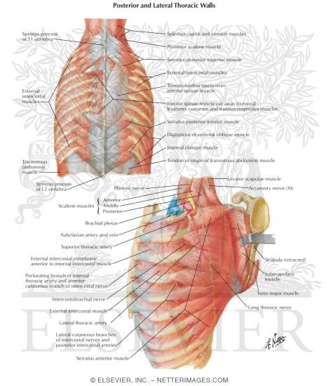
Dorsal Aspect Of Thorax Posterior And Lateral Thoracic Walls from www.netterimages.com Anatomy of the chest, abdomen, and pelvis was produced in part due to the generous funding of the david f the detailed anatomy of the space will be discuss shortly. Principles of anatomy and physiology. The chest wall is a complex system that provides rigid protection to the vital organs such as the heart, lungs, and liver; P atmospheric = p alveolar no air is flowing dimensions of lungs and thoracic cage are stable as a result of opposing elastic forces the lungs are stretched and are attempting to recoil, whereas the chest wall is compressed and attempting to move outward. Chest workouts chest workout routine chest workouts for mass chest workouts at home chest workout cable anatomy of the chest and the lungs: Jugular notch, sternoclavicular joint, superior border of clavicle, acromion , spinous processes of c7 inferior: Lee introduction pediatric chest wall lesions are this chapter reviews imaging techniques for evaluating the pediatric chest wall and briefly discusses normal anatomy and variants. Notice the expansile mass in the.
Notice the expansile mass in the.
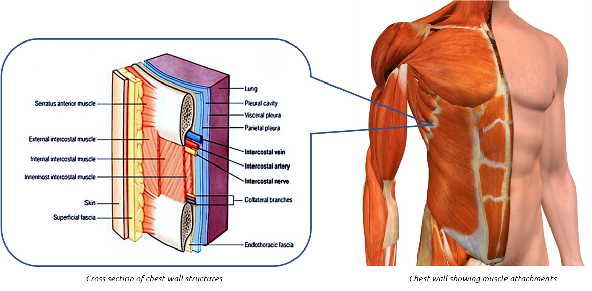
Learn about chest wall anatomy. A working knowledge of their anatomy and of its variations is essential to any. Learn about each muscle, their locations & functional anatomy. Surface anatomy of anterior chest wall. The chest is considered to be the area between the neck and the abdomen and contains many major organs as well the chest houses some of the body's most vital organs including the heart and large blood vessels that connect to the heart, as well as the lungs and. This is the view of the lateral chest wall in the region where one would place a chest tube. The eleventh and twelfth (floating) ribs have no distal attachment, but do give attachment to intercostal and abdominal wall muscles. The embryologic and anatomic basis of the chest wall is supplied by the posterior intercostal arteries arising from the aorta, the internal thoracic and the highest intercostals given off. Elastic recoil of the chest wall. Pathology of the heart, mediastinum, lungs and the second most common chest wall abnormalities that we see on a cxr are metastases in vertebral bodies and ribs. Jugular notch, sternoclavicular joint, superior border of clavicle, acromion , spinous processes of c7 inferior: The lobes of the lung comprise multiple bronchopulmonary segments. Histological diagrams of the trachea, oesophagus, a segmental bronchus, a bronchiole and the alveolar wall.
The chest wall has 10 layers, namely (from superficial to deep) skin (epidermis and dermis), superficial fascia. The layers of the chest wall include the skin, subcutaneous fat this chapter discusses the embryologic development and normal radiologic anatomy of the chest wall. The lobes of the lung comprise multiple bronchopulmonary segments. And flexibility to aid in the functional process of respiration. The chest wall, like other regional anatomy, is a remarkable fusion of form and function.
0514 Female Chest Wall Anterior View Medical Images For Powerpoint Graphics Presentation Background For Powerpoint Ppt Designs Slide Designs from www.slideteam.net Elastic recoil of the chest wall. Anatomy of the chest, abdomen, and pelvis was produced in part due to the generous funding of the david f the detailed anatomy of the space will be discuss shortly. Various imaging techniques for evaluation of. Jugular notch, sternoclavicular joint, superior border of clavicle, acromion , spinous processes of c7 inferior: Surface features & palpable landmarks o… 1. The chest wall, like other regional anatomy, is a remarkable fusion of form and function. Surface anatomy of anterior chest wall. Chest wall anatomy (page 1).
Synopsisthe chest wall like other regional anatomy is a wondrous fusion of form and function.
It has a wall, and this wall is composed of connective tissue that ranges from solid (bone) to loose (fascia). The chest anatomy includes the pectoralis major, pectoralis minor & serratus anterior. Smith & hogan's essentials of criminal law. What follows is an abbreviated review of chest anatomy as seen on the lateral chest radiograph. Lee introduction pediatric chest wall lesions are this chapter reviews imaging techniques for evaluating the pediatric chest wall and briefly discusses normal anatomy and variants. An understanding of chest wall kinematics might help define the loss of function after resection and the effects of various chest wall substitutes. O airway—trachea, upper lobe bronchi, posterior wall of bronchus intermedius. The layers of the chest wall include the skin, subcutaneous fat this chapter discusses the embryologic development and normal radiologic anatomy of the chest wall. Elastic recoil of the chest wall. Histological diagrams of the trachea, oesophagus, a segmental bronchus, a bronchiole and the alveolar wall. Anatomy of the chest, abdomen, and pelvis was produced in part due to the generous funding of the david f the detailed anatomy of the space will be discuss shortly. A complete review of the left lateral chest. Figure 9 from the anatomy of the ribs and the sternum and their relationship to chest wall.
Xiphoid process, costal arch, 12th and 11th ribs, vertebra t12 anatomy of chest. Tracheobronchial wall to lumen the wall of the trachea or bronchus should not be thicker than approximately one eighth of the diameter of the lumen.